Gharbi Classification Hydatid Cyst Treatment : Current Status Of Diagnosis And Treatment Of Hepatic Echinococcosis - After 1 year of albendazole treatment, the cyst had converted to type 2 according to the gharbi classification (ce 3a according to the who classification) (fig.
Gharbi Classification Hydatid Cyst Treatment : Current Status Of Diagnosis And Treatment Of Hepatic Echinococcosis - After 1 year of albendazole treatment, the cyst had converted to type 2 according to the gharbi classification (ce 3a according to the who classification) (fig.. Septated cystic lesion stage 3: 1).then the patient was referred to our interventional department for percutaneous treatment. There was only one case of type iv and no cases of type v. The 2001 world health organization (who) classification of hepatic hydatid cysts is used to assess the stage of hepatic hydatid cysts on ultrasound and is useful in deciding the appropriate management depending on the stage of the cyst. Ultrasonographic appearance of a calcified hydatid cyst.
After percutaneous treatment, the hydatid cysts gradually became solid (gharbi type iv) (figure 1b) and the pain in the muscle resolved. 2 a ct imaging of type 3 cystic hydatid cyst with exophytic distension from the left lobe of the liver, compressing the stomach posteroinferiorly and operative figures of partial pericystectomy in this case (b); 1 treatment algorithms of liver hydatid cysts fig. This classification was proposed by the who in 2001 and, at the time of writing (july 2016), remains the most widely used classification for hepatic hydatid cysts. Type iii has the membrane undulating in the cystic cavity;
Major Hepatic Resection In Hepatic Hydatidosis from www.scirp.org Laparoscopic treatment of hydatid cyst of the liver: The morphologic classifications of lhcs include the sonographic gharbi's classification. Hydatid disease is a worldwide zoonosis produced by the larval stage of the echinococcus tapeworm (, fig 1).the two main types of hydatid disease are caused by e granulosus and e multilocularis.the former is commonly seen in the great grazing regions of the world—particularly the mediterranean region, africa, south america, the middle east, australia, and new zealand—and is. This classification was proposed by the who in 2001 and, at the time of writing (july 2016), remains the most widely used classification for hepatic hydatid cysts. All cysts were classified as per the gharbi's classification. Cyst < 6 cm cyst > 6 cm growth of cyst > 2 cm /peryear growth of cyst < 2 cm /peryear: Gargouri m, ben amor n, ben chehida f, hammou a, gharbi ha, ben cheikh m, et al. After 1 year of albendazole treatment, the cyst had converted to type 2 according to the gharbi classification (ce 3a according to the who classification) (fig.
Septated cystic lesion stage 3:
The appearance at the end of the operation (c) Percutaneous treatment of hydatid cysts (echinococcus granulosus). Hydatid cyst of the liver is the most common clinical presentation of echinococcus granulosus. We report our technique for and results of percutaneous treatment of heterogenous, predominantly solid echopattern hepatic hydatid cysts (hhc), i.e., complex type iv cysts according to gharbi's sonographic classification of hhc. 20 irritating scolicidals such as 95% ethanol are to be avoided in communicating cysts because of the high risk of sclerosing cholangitis. Various classification systems have been published. Percutaneous treatments (pt) and antiparasitic treatment with benzimidazoles (bmz) represent alternatives to surgery. The initial classification by gharbi et al and the world health organization classification are the most commonly preferred. A gharbi type i cyst is a pure fluid collection that is difficult to distinguish from other types of epithelial liver cyst. The liver segments were grouped as near to the hilum (segments i, iii, ivb, v, and vi) and remotely distant (segment ii, iva, vii, and viii) with modification of classification by dziri et al. Doppler ultrasonography is indicated to show the reports of hydatid cyst with vascular axes (portal vein, hepatic veins, and inferior vena cava). Antibiotics are used prophylactically for surgery as indicated in patients with a cystobiliary fistula, for the treatment of infected cysts, and for the treatment of associated infections. Spleen and kidneys are the organs where hydatid disease is most frequently observed after the liver and lung.
Type iii hydatid cysts are those with fluid collection and septa. The gharbi classification system was used to stage the hydatid disease 9 . The liver segments were grouped as near to the hilum (segments i, iii, ivb, v, and vi) and remotely distant (segment ii, iva, vii, and viii) with modification of classification by dziri et al. Communicating cysts are generally unsuitable for percutaneous treatment because cyst fluid, hydatid sand and scolicidal agents are forced into biliary radicles as a result of increased intracyst pressure. However, in the types i and iv, we have to consider differential diagnosis.
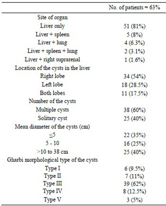
Major Hepatic Resection In Hepatic Hydatidosis from www.scirp.org There was only one case of type iv and no cases of type v. All cysts were classified as per the gharbi's classification. According to gharbi's classification, three cases (21.4 %) of the unusually located hydatid cysts were type i, two (14.3 %) type ii, and eight (57.1 %) type iii. There are several classification schemes for liver hydatid cysts based on their ultrasound appearances; Septated cystic lesion stage 3: The morphologic classifications of lhcs include the sonographic gharbi's classification. Doppler ultrasonography is indicated to show the reports of hydatid cyst with vascular axes (portal vein, hepatic veins, and inferior vena cava). The cysts should be larger than 5 cm in diameter and type i or ii according to the gharbi ultrasound classification of liver cysts (ie, type i is purely cystic;
After percutaneous treatment, the hydatid cysts gradually became solid (gharbi type iv) (figure 1b) and the pain in the muscle resolved.
20 irritating scolicidals such as 95% ethanol are to be avoided in communicating cysts because of the high risk of sclerosing cholangitis. Antibiotics are used prophylactically for surgery as indicated in patients with a cystobiliary fistula, for the treatment of infected cysts, and for the treatment of associated infections. The appearance at the end of the operation (c) Doppler ultrasonography is indicated to show the reports of hydatid cyst with vascular axes (portal vein, hepatic veins, and inferior vena cava). The patient was followed up for almost eight years with ultrasonography and ct. This is particularly the case in the early cyst stages when hydatid fluid is still tightly contained within the endocysts (cyst stage ce1) and in the final stage of involution (ce5) when cyst content is solid and the cyst wall largely calcified. Treatment of gharbi type iii hydatid cysts is still controversial. This classification was proposed by the who in 2001 and, at the time of writing (july 2016), remains the most widely used classification for hepatic hydatid cysts. We report our technique for and results of percutaneous treatment of heterogenous, predominantly solid echopattern hepatic hydatid cysts (hhc), i.e., complex type iv cysts according to gharbi's sonographic classification of hhc. There was only one case of type iv and no cases of type v. Type of cyst according to gharbi classification : There are several classification schemes for liver hydatid cysts based on their ultrasound appearances; Hydatid cyst of the liver is the most common clinical presentation of echinococcus granulosus.
All cysts were classified as per the gharbi's classification. This is particularly the case in the early cyst stages when hydatid fluid is still tightly contained within the endocysts (cyst stage ce1) and in the final stage of involution (ce5) when cyst content is solid and the cyst wall largely calcified. Ultrasonographic appearance of a calcified hydatid cyst. There are several classification schemes for liver hydatid cysts based on their ultrasound appearances; A gharbi type i cyst is a pure fluid collection that is difficult to distinguish from other types of epithelial liver cyst.
Gharbi Classification Of Hydatid Cysts 6 Download Scientific Diagram from www.researchgate.net Discover (and save!) your own pins on pinterest Gargouri m, ben amor n, ben chehida f, hammou a, gharbi ha, ben cheikh m, et al. Calcified or partially calcified lesion (inactive cyst) treatment and prognosis According to gharbi's classification, three cases (21.4 %) of the unusually located hydatid cysts were type i, two (14.3 %) type ii, and eight (57.1 %) type iii. However, in the types i and iv, we have to consider differential diagnosis. A catheterization technique was performed but hypertonic saline and alcohol were not given into the cavity due to cystobiliary leakage. This is particularly the case in the early cyst stages when hydatid fluid is still tightly contained within the endocysts (cyst stage ce1) and in the final stage of involution (ce5) when cyst content is solid and the cyst wall largely calcified. Type of cyst according to gharbi classification :
However, in the types i and iv, we have to consider differential diagnosis.
Percutaneous treatments (pt) and antiparasitic treatment with benzimidazoles (bmz) represent alternatives to surgery. However, in the types i and iv, we have to consider differential diagnosis. 20 irritating scolicidals such as 95% ethanol are to be avoided in communicating cysts because of the high risk of sclerosing cholangitis. This is particularly the case in the early cyst stages when hydatid fluid is still tightly contained within the endocysts (cyst stage ce1) and in the final stage of involution (ce5) when cyst content is solid and the cyst wall largely calcified. There are several classification schemes for liver hydatid cysts based on their ultrasound appearances; Calcified or partially calcified lesion (inactive cyst) treatment and prognosis Spleen and kidneys are the organs where hydatid disease is most frequently observed after the liver and lung. 2 a ct imaging of type 3 cystic hydatid cyst with exophytic distension from the left lobe of the liver, compressing the stomach posteroinferiorly and operative figures of partial pericystectomy in this case (b); Type iii hydatid cysts are those with fluid collection and septa. Polat fr, polat s, sultanoglu e. Cystic lesion with daughter lesions; Type ii is purely cystic plus hydatid sand; Laparoscopic treatment of hydatid cyst of the liver:


0 Komentar